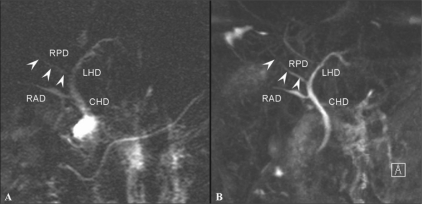
Figure 16 (A,B).
A 35-year-old man with an anatomical variation of the biliary tree. 2D coronal T2W TSE image (TR 2800, TE 1100, FA 150) (A) shows drainage of the RPD (arrowheads) into the left main duct with separate drainage of the RAD into the CHD. 3D coronal T2W (TR 4235, TE 545, FA 180) MIP image (B) demonstrates this with better resolution. RPD: right posterior duct, RPD (arrowheads) is more clearly depicted on 3D MIP image. RAD: right anterior duct, LHD: left hepatic duct, CHD: common hepatic duct

