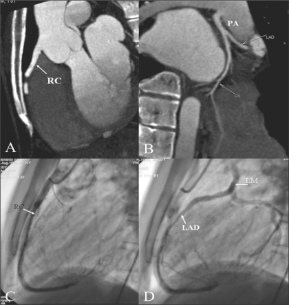
Figure 12 (A–D).
ALCAPA. Sagittal MIP image igure (A) shows normal origin of the RCA (RC) from the right cusp. MIP image (B) reveals the LM originating from the PA — ALCAPA. Lateral coronary angiogram (C) shows a normal RCA (RC). Late phase of the same run (D) shows retrograde filling of the CX, LAD, and then the LM, which eventually opens into the PA

