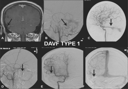
Figure 1: (A-F).
DAVF Type 1: Postcontrast coronal T1W MRI image (A) shows multiple tortuous flow voids (arrow) adjacent to the right sigmoid sinus. Selective right external carotid artery (ECA) (B) and internal carotid artery (ICA) (lateral view) (C) angiograms shows a DAVF type 1 with feeders (arrow) from the posterior meningeal branch of the middle meningeal artery and dural branches (arrow) from the cavernous ICA draining antegradely through the sigmoid sinus. Posttreatment selective right ECA (lateral view) (D) and ICA (anteroposterior view; arterial (E) and venous (F) phases) angiograms show coils (arrows) packing the right distal transverse and sigmoid sinus with total obliteration of the fistula

