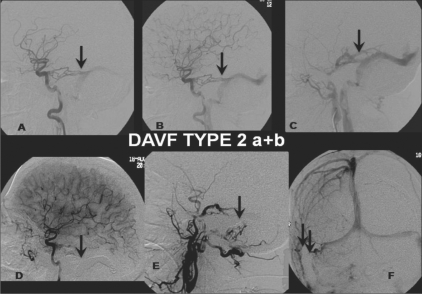
Figure 2: (A-F).
DAVF Type 2 a + b: Selective right ICA (A, B) and ECA (lateral view) (C) angiograms show a DAVF type 2 a + b, with feeders from the dural branches (arrow) of the ICA and the posterior meningeal branch of the middle meningeal artery (arrow) draining retrogradely through the transverse sinus. Post-treatment selective right ECA (lateral view) (D) and ICA (lateral view, arterial phase (E); anteroposterior view, venous phase (F)) angiograms show coils (arrows) packing the right distal transverse and sigmoid sinus with total obliteration of the fistula

