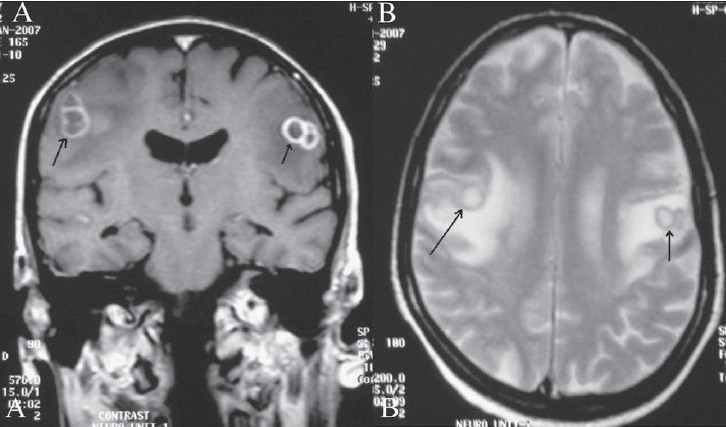
Figure 1 (A, B).

Contrast T1W coronal (A) and axial T2W MRI images of the brain show conglomerate ring-enhancing lesions in both the frontal subcortical regions (arrows in A), with central hyperintensity and peripheral low signal intensity rims (arrows in B)
