Figure 5 (A, B).

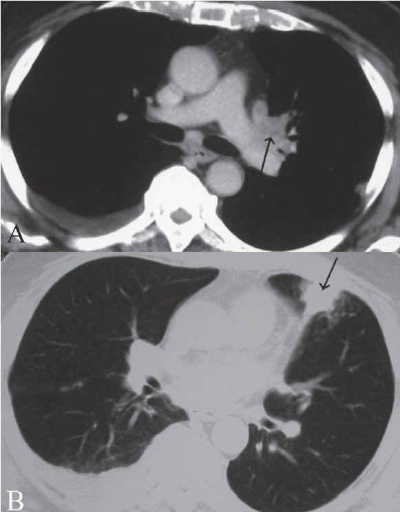
Soft tissue (A) and lung window (B), contrast-enhanced CT images of the chest show necrotic lymphnodes in the left hilar region (arrow) with consolidation in the left lung (arrow)

Soft tissue (A) and lung window (B), contrast-enhanced CT images of the chest show necrotic lymphnodes in the left hilar region (arrow) with consolidation in the left lung (arrow)