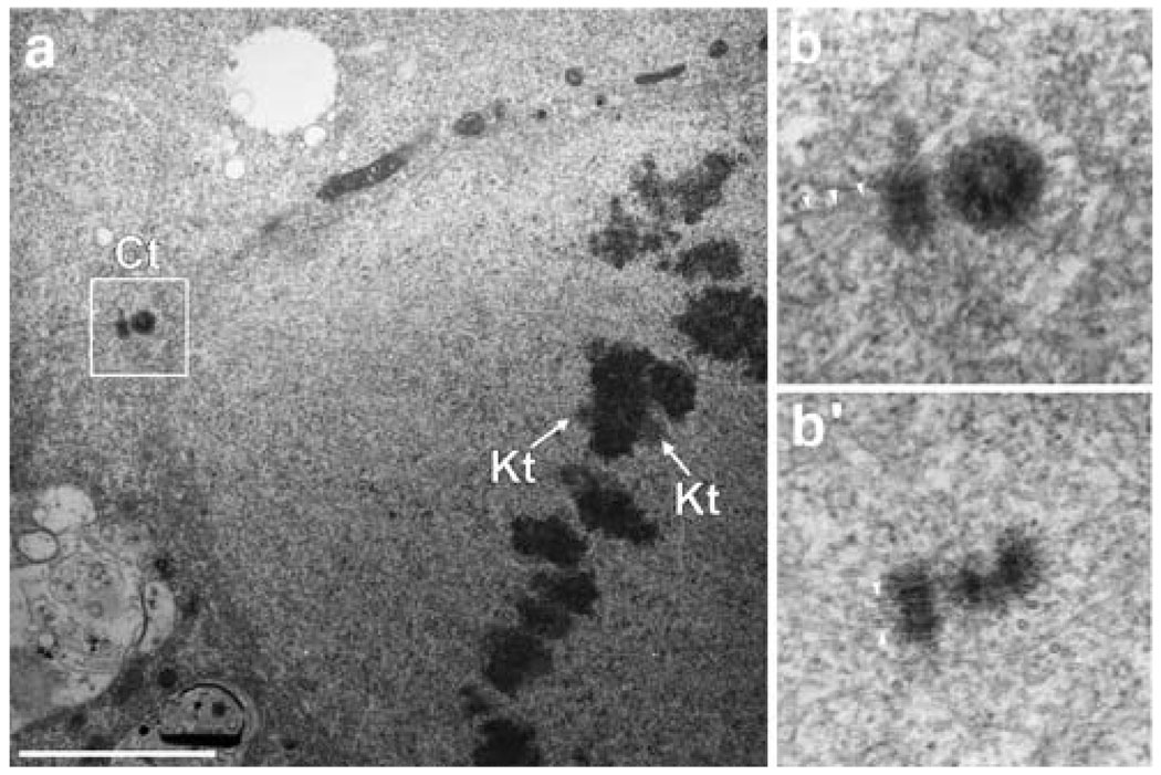
Fig. 3.
Ultrastructural characterization of the mitotic apparatus of Drosophila S2R+ cells. a. Visualization of a half-spindle region from S2R+ cells showing one centrosome (Ct) composed of two centrioles, plus a kinetochore pair (Kt) with attached MTs. Several K-fibers can be identified. b Higher-magnification view of the centrosome visualized in a. b′. Similar view of the other centrosome. Note that both centrioles have MTs directly attached (arrowheads). Scale bar=2.5 µm

