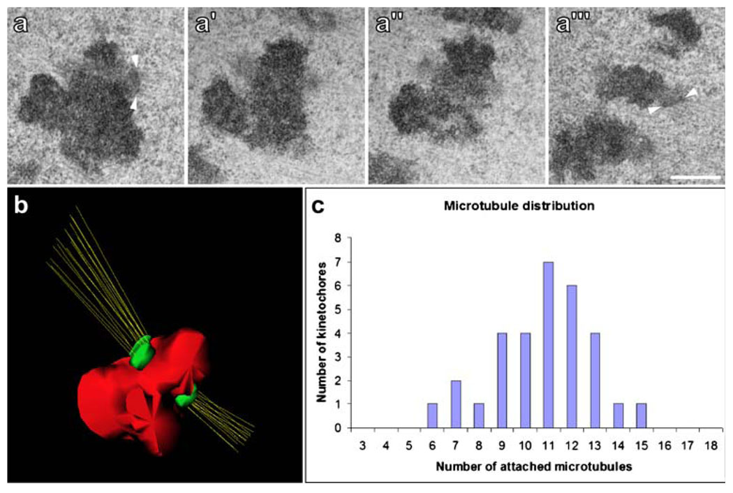
Fig. 4.
The Drosophila mitotic kinetochore and K-fiber. a–a''' Serial-sections through a kinetochore from a metaphase S2R+ cell. Note the highly amorphous structure that protrudes at the centromeric region. In some favorable views, such as a and a''', it is possible to visualize a thin electron-dense layer immediately adjacent to the amorphous material where MTs are bound (arrowheads). Scale bar=0.5 µm. b Three-dimensional reconstruction from serial sections of the chromosome observed in a. In this particular case, the sister kinetochores are bound to 15 and 12 MTs. c Direct MT counts from 31 kinetochores. The average number of kMTs bound is 10.8 MTs/kinetochore, with a standard deviation of 2.1, and a range of 6–15

