
Figure 7. Summary model comparing the vascularization of adult and embryonic germinal zones.
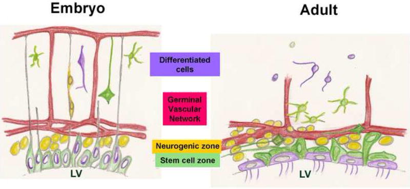
In the embryo, an apical layer of stem cells produces an actively proliferating SVZ which generates neurons that migrate towards the pia guided by radial glia. Coincident with neurogenesis, the vasculature grows from the pial surface towards the germinal cells, a source of VEGF (Breier et al., 1992) and form a plexus at the germinal zones, parallel to the ventricular surface (Shen et al., 2004; Strong, 1964).
The adult SVZ has an essentially similar structure: Apical Type B cells, believed to include the SVZ stem cells, are intercalated into the ependymal layer and directly contact the ventricle. Just subjacent is an SVZ of active proliferation, differentiation and migration, including Mash1+ and Olig2+ Type C progenitors, PSA-NCAM+ Type A neuroblasts and Tangential Type B cells. The adult germinal zone is intimately associated with an SVZ vascular plexus.