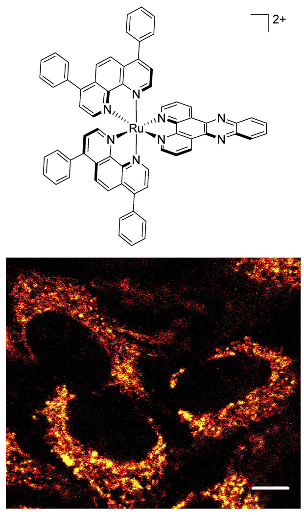
Figure 1.
A luminescent ruthenium probe used to examine metal complex uptake. Top: Chemical structure of Ru(DIP)2dppz2+. Bottom: HeLa cells incubated with 5 µM Ru(DIP)2dppz2+ for 4 h, imaged by confocal microscopy. Note that the cytoplasm is extensively stained with the Ru complex. Scale bar is 10 µm.

