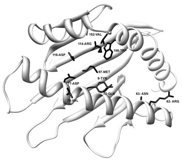
Figure 2.

Structure of HLA*6801. Amino acids that are observed in the largest number of different mismatch combinations are distributed throughout the peptide binding domain. The side chains of the amino acids that are observed in the largest number of different mismatch combinations are shown on an HLAA*6801 structure (2HLA in the Protein Data Bank). Molecular graphics images were produced using the UCSF Chimera package from the Resource for Biocomputing, Visualization, and Informatics at the University of California, San Francisco.
