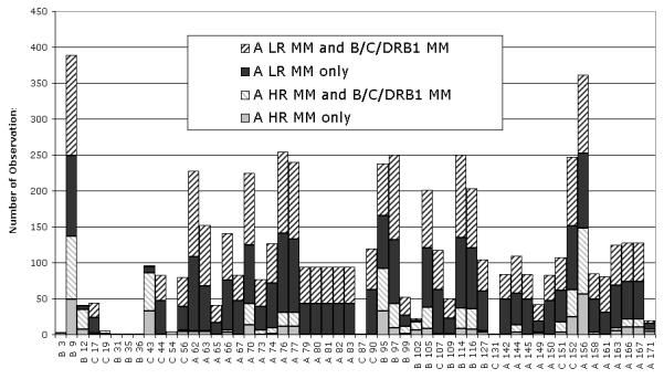
Figure 3.

Amino acid differences that are involved in HLA-A disparities are distributed throughout the peptide binding domain, present in both low resolution (LR) and high resolution (HR) mismatches, and often accompanied by mismatches (MM) at HLA-B, -C, and DRB1 loci. The position and side-chain orientation of each mismatched amino acid are indicated below the x-axis (A=alpha helix, B= beta sheet, and C=connecting loop). The amino acids that are most frequently mismatched line the peptide binding groove of the HLA molecule. Most of the mismatched amino acids are involved in both low and high resolution mismatches.
