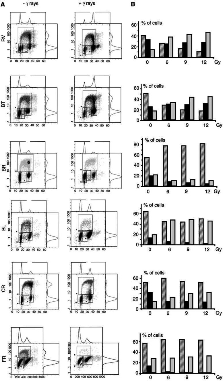
Figure 1.
Flow cytometric analysis of HMCLs after exposure to γ-radiation. (A) Left: nonirradiated cells; right: 24 h after exposure to 6 Gy. Double labelling: PI (x-axis) and BrdU (y-axis). The different areas N, M and P represent cells in G0/G1, cycling S cells and cells in G2-M, respectively. (B) Dose-dependent cell distribution in different phases of the cell cycle. Percentage of cells in G0/G1 (hatched bars), S (black bars), G2/M (grey bars), 24 h after exposure to 6, 9 and 12 Gy.

