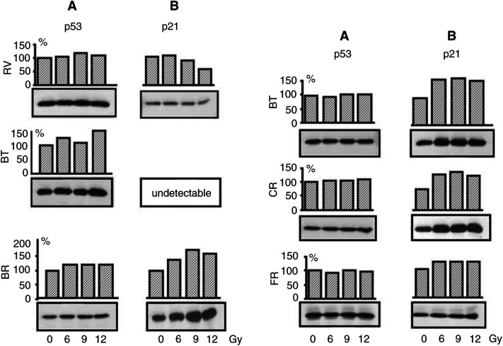
Figure 3.
p53 (A) and p21WAF1/CIP1 (B) protein expression in HMCLs after exposure to several doses of γ-radiation. At 24 h after irradiation, protein extracts were subjected to SDS-PAGE electrophoresis followed by immunoblot analysis with antibodies against the corresponding antigens. ECL detection. Densitometric analyses of p53 and p21WAF1/CIP1 expression are reported on the top of the corresponding bands as percentage of the amount of protein expressed in untreated cells.

