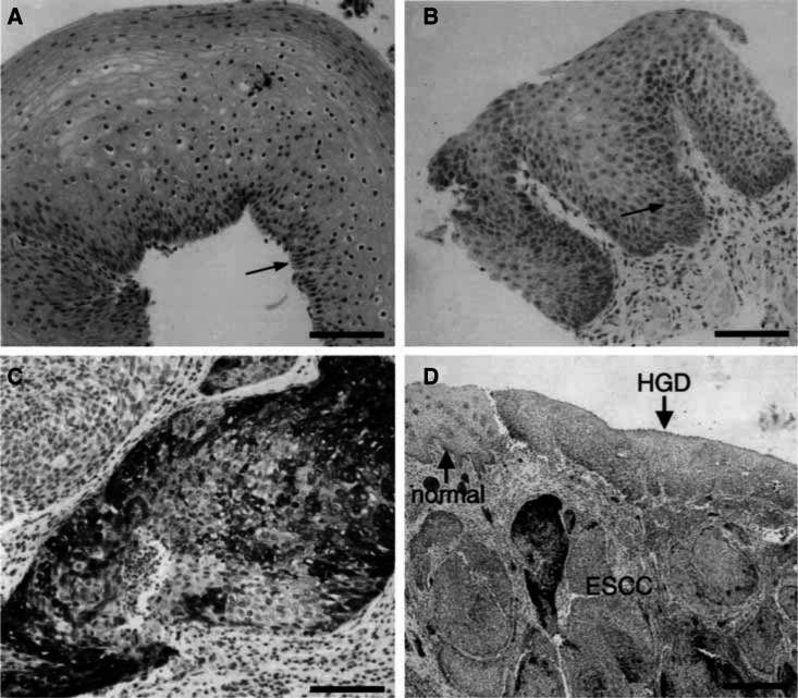
Figure 2.
COX-2 immunoreactivity in normal and neoplastic oesophageal squamous tissues of HNC patients. (A) Normal oesophageal squamous epithelium exhibits COX-2-specific staining only in cells of the basal layer (arrow); the staining score is 1. (B) Squamous cells of LGD demonstrate moderate COX-2-specific staining; the staining score is 4. (C) Poorly differentiated ESCC shows heterogeneous COX-2 staining; the staining score is 4. (A–C) Bar=100 μM. (D) Resected oesophageal tissue showing COX-2 expression that increases from normal squamous mucosa (normal) to HGD and to carcinoma (ESCC). Bar=500 μM.

