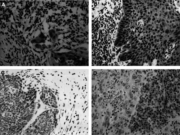
Figure 1.
Immunohistochemical staining of Fhit protein in cervical cancer. (A, B) Normal Fhit expression in cervical cancer. The Fhit expression scores were 6 (A) and 9 (B). (C) Absent Fhit expression in cervical cancer. Fhit expression in the cervical cancer tissue was negative (score 0). Note that Fhit is expressed normally in the normal stromal tissue. (D) Marked reduced Fhit expression in cervical cancer. The Fhit expression score in cancer tissue was 3.

