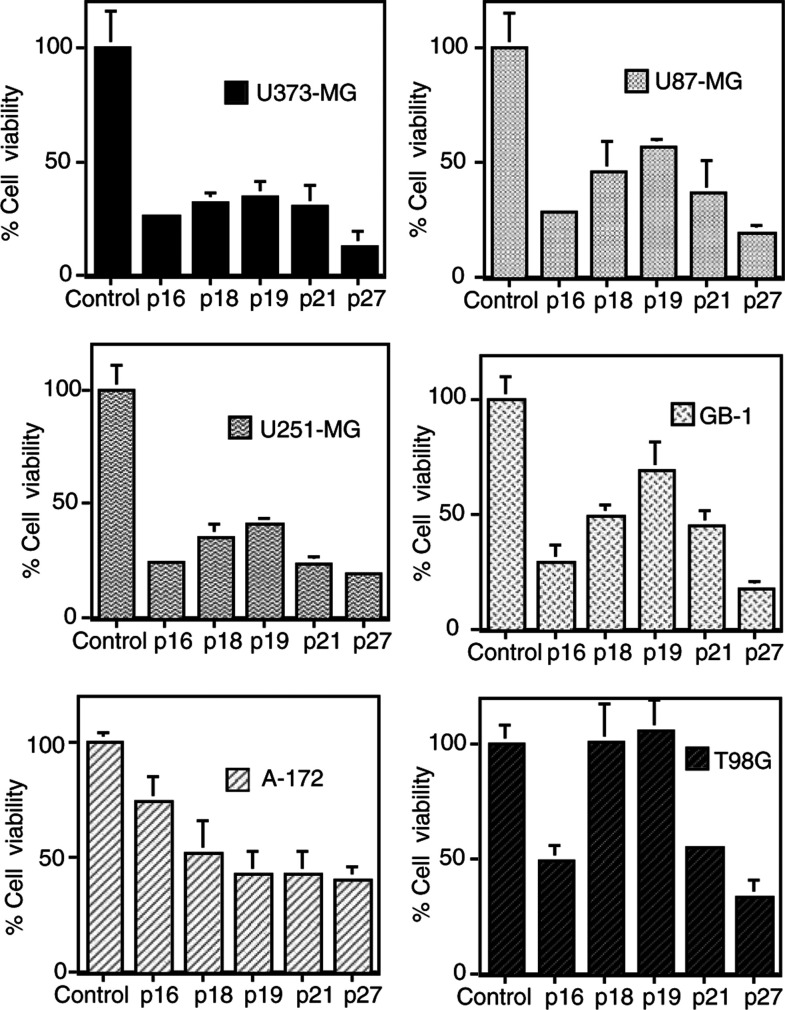
Figure 1.
Effect of adenovirus expressing CDKIs on cell viability of malignant glioma cell lines. Tumour cells were seeded at 5 × 10³ cells well−1 (0.1 ml) in 96-well flat-bottomed plates and incubated overnight at 37°C. On the following day, U373-MG, U251-MG, U87-MG cells, A172 cells, GB-1 and T98G cells were infected with AdBHGΔl,3, AdMH4pl6, AdMH4pl8, AdMH4pl9, AdMH4p21 or AdMH4p27 as indicated (day 0). On day 3, the cell viability was determined using a trypan blue dye exclusion assay. Values are given as the percentage of viable cells of AdBHGΔl ,3-infected cultures. Results shown are the means±s.d. of three independent experiments. U373-MG, U251-MG, U87-MG, A172 and GB-1 cells were infected at 60 MOI, while 180 MOI was used for T98G cells.

