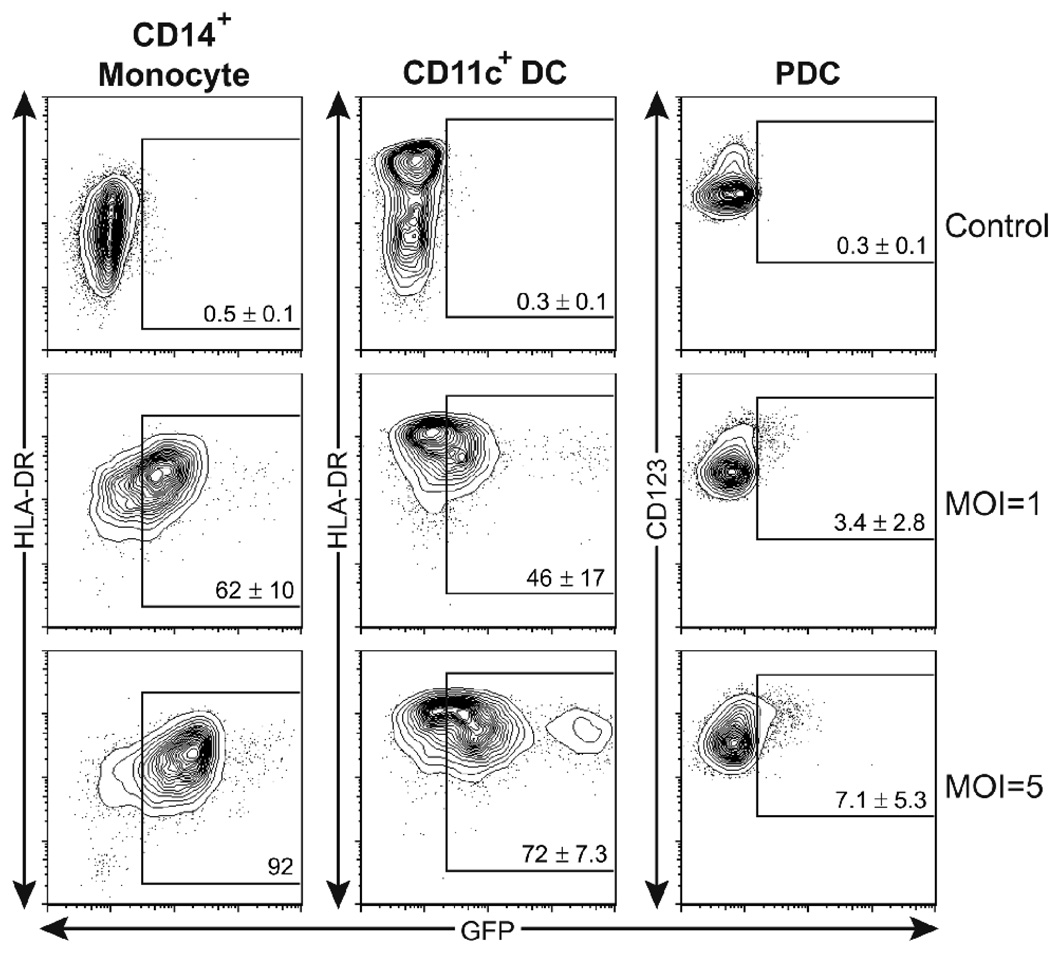
Fig. 1. HCMV targets monocytes and myeloid DCs in peripheral blood.
Freshly isolated blood CD14+ monocytes (left panels), CD11c+ DCs (middle panels) and CD123+ PDCs (right panels) were infected for 36 h with GFP-recombinant HCMV at MOI’s of 1 and 5. The expression of indicated surface markers and of GFP identifying HCMV-infected cells was assessed by FACS. Shown are 5% contour plots with outliers of cells after gating leukocytes via FSC–SSC and CD14+to identify monocytes, FSC–SSC and CD11c+ to identify CD11c+ DCs, and FSC–SSC and CD11c–/CD123+ to identify PDCs. The percentage of GFP+ cells, among the identified cell populations is shown in each plot, indicated by a box. Numbers are presented as the mean values ± SD of data from 3–4 individual donors.

