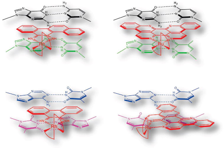
Figure 4.

Schematic illustrations of Ru(bpy)2(eilatin)2+ (right) bound to mismatched (top) and matched (bottom) DNA sites based on the crystal structures of chrysi (top left) and phi (bottom left) complexes of Rh bound to mismatched and matched DNA, respectively. For binding to the mismatched site, the metal complexes are oriented from the minor groove side, whereas for binding to the matched site, the association is from the major groove side.
