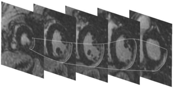
Figure 1. Stack of short axis DE-MRI pictures showing delayed enhancement in the epicardial left ventricular lateral wall.

Although the free wall is thinned, the endocardium is relatively preserved. The epicardial contours of the scar are traced in dotted lines. An endocardial radiofrequency catheter ablation procedure for VT was unsuccessful. Epicardial ablation successfully eliminated all 3 inducible VTs.
