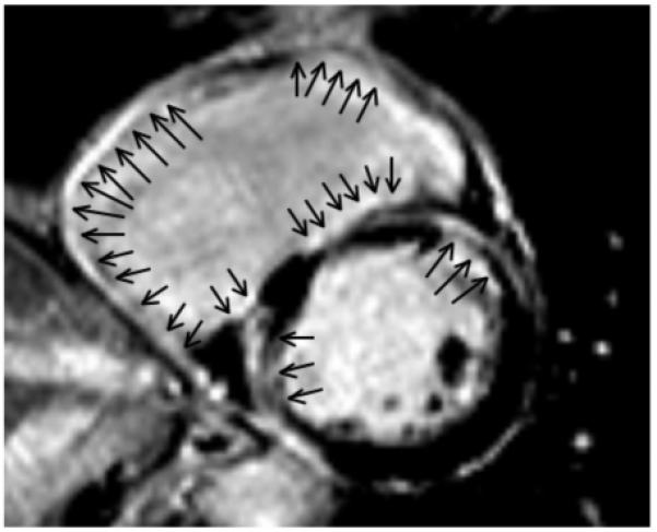
Figure 3. A short-axis view of the mid-portion of the right and left ventricles.

The scar involves predominantly the right ventricular endocardium. The right ventricle shows enodcardial delayed enhancement (arrow) that is transmural in the right ventricular free wall and mostly endocardial at the right ventricular septum. Endocardial scar also is present in the left ventricle, with some transmural components. All 4 inducible VTs were ablated at the right ventricular endocardium in this patient.
