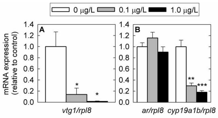
Fig. 5.
Gene expression changes in female fathead minnow liver (A) and brain (B) measured by Q-PCR following exposure to 17β-trenbolone for 4 days. A) vitellogenin 1 (vtg1) in liver tissue. B) cytochrome P450, family 19, subfamily A, polypeptide 1b (cyp19a1b) and androgen receptor (ar) in brain tissue. mRNA expression was normalized to the abundance of ribosomal protein L8 (rpl8). Experimental groups consisted of six fish (liver) or twelve fish (brain) and each fish was analyzed in duplicate. Error bars denote the standard error of the mean. Statistically significant differences in expression levels between control and treated fish are denoted by stars (* P < 0.05; ** P < 0.01; *** P < 0.001).

