
Fig. 2.
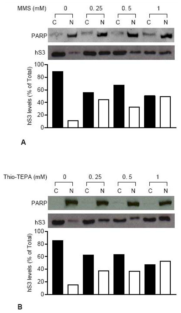
Nuclear translocation of hS3 in response to MMS and Thio-TEPA treatment. Cytosolic (C) and nuclear (N) fractions were prepared from HEK 293 cells exposed to various concentrations of MMS (A) or Thio-TEPA (B) for 24 h. After immunoblotting analysis of fractions using anti-hS3 antibody, sub-cellular levels of hS3 were evaluated from integrated density values, obtained by densitometry tracing of each band. Cytosolic and nuclear density values for each sample were added to obtain total cellular hS3 and hS3 levels in individual fractions are represented as percent of total hS3. Localization of nuclear protein PARP on both blots was traced by stripping and re-probing with anti-PARP antibody.