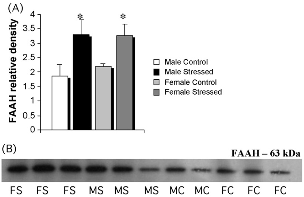
Fig. 5.
Stress increases FAAH in both males and females in dorsal hippocampus. (A) Tissue from the same animals in Fig. 4 was also assessed for levels of FAAH protein by western blot. There was a significant increase in FAAH in both males and females after three weeks of unpredictable stress (two-way ANOVA; p < 0.03). (B) Representative photomicrographs of western blots showing visualization of bands for FAAH: FS = female stressed, FC = female control, MS = male stressed, MC = male control.

