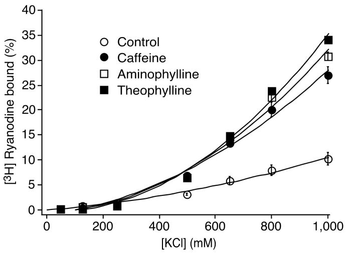
Fig. 7. Effects of methylxanthines on the basal activity of [3H]ryanodine binding.
[3H]ryanodine binding to cell lysate prepared from HEK293 cells expressing RyR2 was carried out at ~3 nM Ca2+ (pCa 8.49), various concentrations of KCl (50–1000 mM), 2.5 mM caffeine (filled circles), aminophylline (open squares) or theophylline (filled squares), and 5 nM [3H]ryanodine. The channel activity in the absence of methylxanthines (control) is also shown (open circles). The amount of [3H]ryanodine binding at various Ca2+ concentrations was normalized to the maximal binding at 1000 mM KCl and pCa 4. Data points shown are mean ± SEM from 3 separate experiments.

