
FIGURE 6.
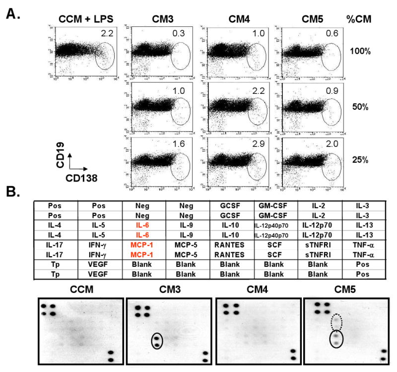
MSCs prevent B cell differentiation into plasma cells by releasing humoral factor(s): (A) 106 B cells were cultured with 3 different concentrations (100, 50 or 25%) of CM3, CM4 or CM5 in 24-well plates for 3 days. CMs containing LPS were diluted with LPS-containing CCM, and those without LPS were diluted with CCM. B cells cultured with CCM containing LPS were used as controls. The numbers in each oval indicate the percentages of CD138+ CD19+/- cell fractions (n=3); (B) The presence of cytokines/chemokines in CM3, CM4, and CM5 was screened using the RayBio Mouse Cytokine Array I. The layout of the array is shown on the top. The protein array was incubated separately with test CM and CCM as a control. The results are shown at the bottom. Solid dots indicate the signal for MCP-1 and the circled dots indicates the signal for IL-6. (Pos: positive control; Neg: negative control; Tp: thrombopoietin). Spot intensity relative to the positive and negative control offers an indication of the relative amount of chemokines/cytokines present in the CM.