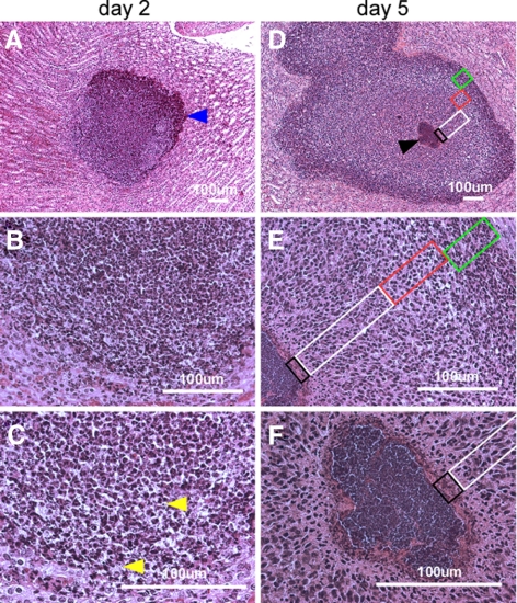
Figure 2.
Histopathology of SACs. BALB/c mice were infected with S. aureus Newman via retro-orbital injection. Thin-sectioned, H&E-stained tissues of infected kidneys on d 2 (A–C) and d 5 following infection (D–F) were analyzed by light microscopy, and images were captured. On d 2, a massive infiltrate (A, blue arrowhead) of PMNs with occasional intracellular staphylococci (C, yellow arrowheads) are characteristic of early infectious lesions. By d 5, staphylococcal abscess communities developed as a central nidus (D, black arrowhead). Staphylococci were enclosed by an amorphous, eosinophilic pseudocapsule (black box) and surrounded by a zone of dead PMNs (white box), a zone of apparently healthy PMNs (red box), and a rim of necrotic PMNs (green box), separated through an eosinophilic layer from healthy kidney tissue.

