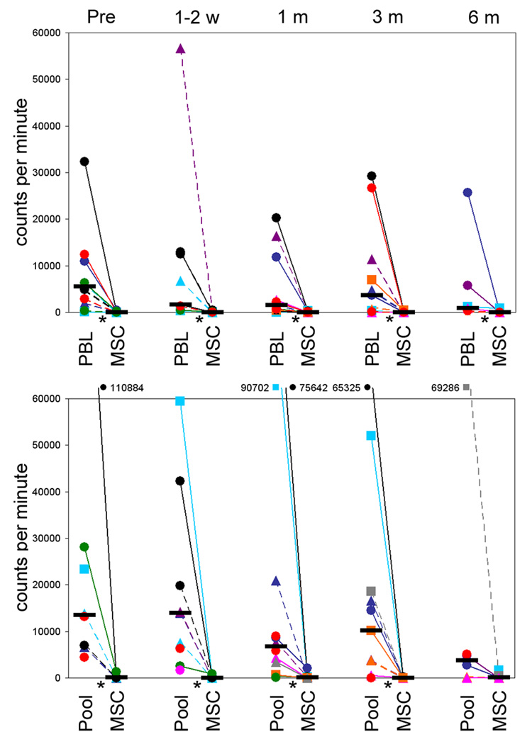
Figure 1.
MSC recipient lymphocyte proliferative responses to MSC donor PBL and MSC (upper panel) and to a pool of allogeneic PBL and to third party MSC (lower panel); at pre-MSC infusion (pre), 1–2 weeks (1–2 w), 1 month (1 m), 3 months (3 m) and 6 months (6 m) post-MSC infusion. Unfractionated PBL were cultured in microtitre plates with irradiated PBL (ratio 1:1) or MSC (ratio 10:1) for 6 days. The MSC were derived from HLA identical siblings (■), or from haploidentical (▲) and mismatched donors (●). Data shown for UPN: 1118, black plain; 1068, red plain; 1044, blue plain; 1115, green plain; 995, light-blue plain; 1098, purple plain; 1110, orange plain; 1047, pink plain; 1126, gray plain; 981, black dashed; 1007, red dashed; 924, blue dashed; 1082, green dashed; 917, light-blue dashed; 868, purple dashed; 994, orange dashed; 1033, pink dashed; 1020, gray dashed (see table 2 for details). Horizontal black bar denotes median proliferation and statistical significance (p<0.05) is indicated by *.

