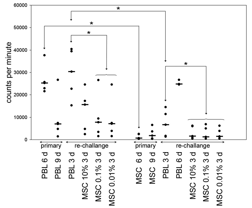
Figure 2.
Lymphocyte proliferation to PBL and MSC in primary and rechallenge co-cultures. Responder PBL were cultured with irradiated, allogeneic PBL or MSC from the same donor for 6–9 days. In restimulation experiments responder PBL were resuspended and 10×105 cells transferred to microtitre plates and rechallenged with PBL or MSC from the original stimulator for 3 days. Proliferation to PBL peaked at 6 days in primary culture and at 3 days on rechallenge (columns 1–3). When responders were rechallenged with MSC from the same donor there was only modest proliferation at 3 days to the highest MSC concentration (columns 3–6). When MSC were used as primary challenge there was minimal proliferation compared with PBL (columns 7–8) and these MSC primed responders mounted a proliferative response to corresponding PBL that followed primary challenge kinetics, i.e. maximum at 6 days, (columns 9–10). Similarly, responders primed with MSC failed to proliferate on rechallenge with MSC (columns 11–13). Horizontal black bars denote median proliferation and statistical significance (p<0.05) is indicated by *.

