FIG. 2.
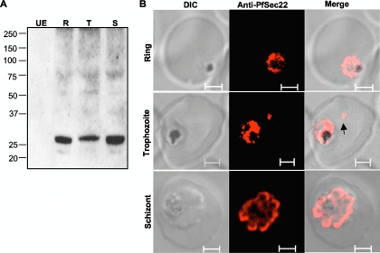
Expression analyses of PfSec22. Antibodies were generated against a peptide sequence in the nonconserved region of PfSec22 and affinity purified as described in Materials and Methods. (A) Immunoblot analysis of parasite extracts showing expression of endogenous PfSec22 in ring (R), trophozoite (T), and schizont (S) stage parasites. Absence of antibody reaction with the uninfected erythrocyte lysate (UE) indicates high specificity of the antibodies for the parasite protein. (B) Immunofluorescence microscopy of fixed cells using anti-PfSec22 antibodies and goat anti-rabbit Alexa Fluor 555 secondary antibodies. An intense ring of PfSec22 fluorescence is visible in ring- and trophozoite-infected cells. Isolated foci of PfSec22 fluorescence (arrow) are also detected in the host cell compartment in trophozoite-infected cells, suggesting export of PfSec22 into the erythrocyte cytosol. Scale bars, 2 μm.

