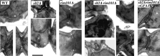
FIG. 3.
Ultrastructure of the yeast septum. TEM images of cells showing the septum structure in different strains. Note the more prominent scars (*) in the slt2Δ rim101Δ double mutant compared to those in the wt or slt2Δ strains and the irregular shape of the primary septum, observed as a white region between septal walls (arrowheads). Also note the more tubular aspect of the neck in the double mutant. The bar corresponds to 0.5 μm.

