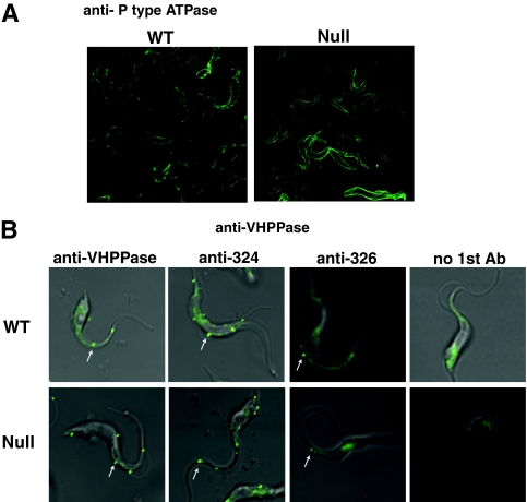
FIG. 7.
Localization of P-type ATPase and VHPPase in wild-type and TbGPI12 null procyclic cells by immunofluorescence microscopy. (A) Fixed and permeabilized cells were stained with rabbit anti-P-type ATPase antibodies followed by fluorescently labeled secondary antibodies, revealing cell surface labeling in both cases. (B) Live cells were incubated with no primary antibody (no 1st Ab; right panels), antibodies raised to a fragment of recombinant VHPPase antibodies (left panels), or one of two anti-VHPPase synthetic peptide antibodies (middle panels); fixed; and then stained with fluorescently labeled secondary antibodies. Punctate staining of surface structures is indicated by white arrows.

