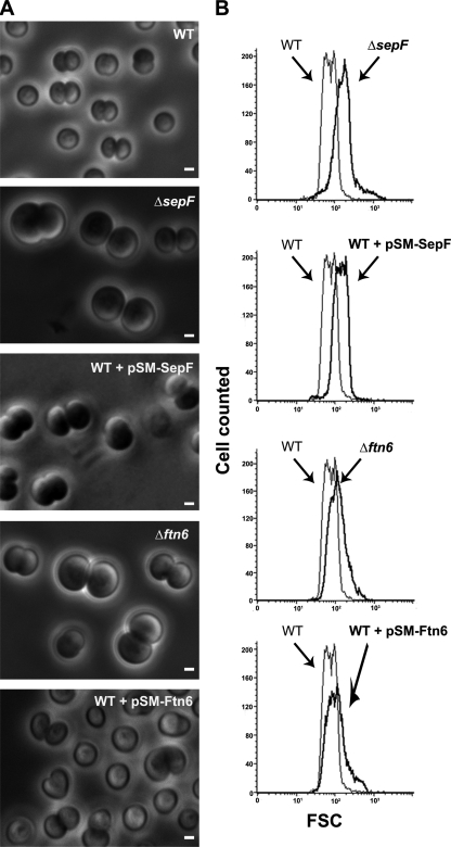
FIG. 1.
Influence of the abundance of SepF and Ftn6 proteins on the morphology and size of Synechocystis cells. Phase-contrast image (A) (scale bars, 1 μm) and flow cytometry analysis (B) of WT or mutant cells depleted with respect to (Δ) or producing the natural or tagged versions of the studied proteins from a pSM expression plasmid (Table 1). For each mutant strain, the FSC histogram (bold lines) has been overlaid with that of the WT to better visualize the influence of the mutation on cell size. These experiments were performed twice with two independent clones harboring the same mutation.

