
FIG. 5.
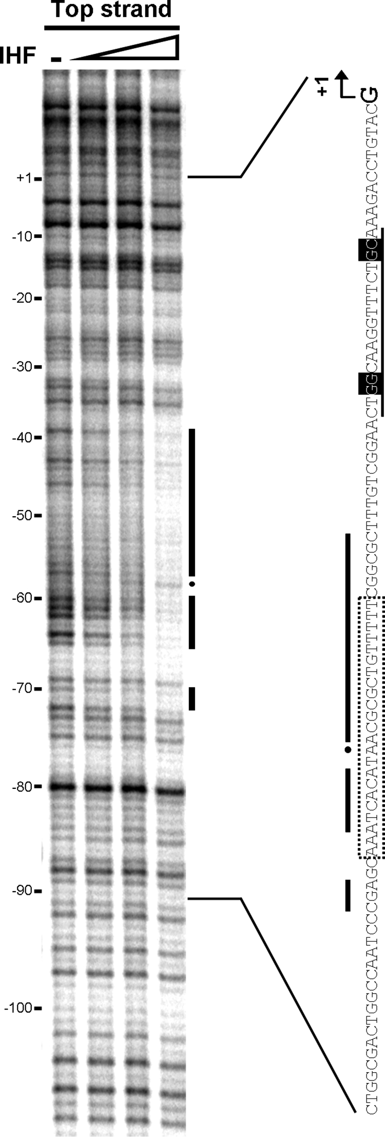
IHF DNase I footprint of the glnK promoter region. The coordinates are relative to the glnK transcriptional start site. IHF concentrations were 0.5, 1, and 2 μM of dimer. The protected regions (black bars) and hypersensitive positions (dots) are marked in the footprint and on the promoter sequence beside it. The σN promoter and the putative IHF binding site identified by sequencing are also marked on the sequence, as described in the legend for Fig. 2.