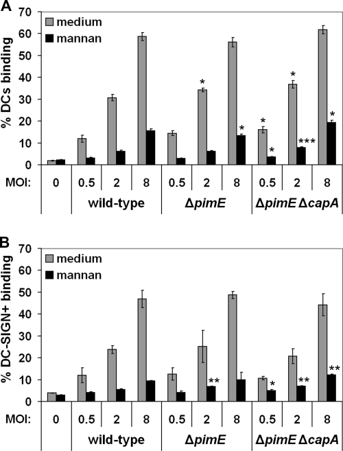
FIG. 5.
Binding assay with (A) MoDCs and (B) Raji cells expressing DC-SIGN. Cells were incubated with wild-type M. bovis BCG or with the ΔpimE and ΔpimE ΔcapA mutants at a MOI of 0.5, 2, or 8 for 45 min in the presence or absence of mannan. The percentage of the cell population binding M. bovis BCG was determined by flow cytometry. Shown are means of triplicates and the standard deviations. For binding to both MoDCs and Raji + DC-SIGN cells, one representative experiment out of three is shown. See Fig. S3 in the supplemental material for the control binding assay with wild-type Raji cells. ***, P < 0.0005; **, P < 0.005; *, P < 0.05 (compared to wild-type M. bovis BCG at equal MOIs).

