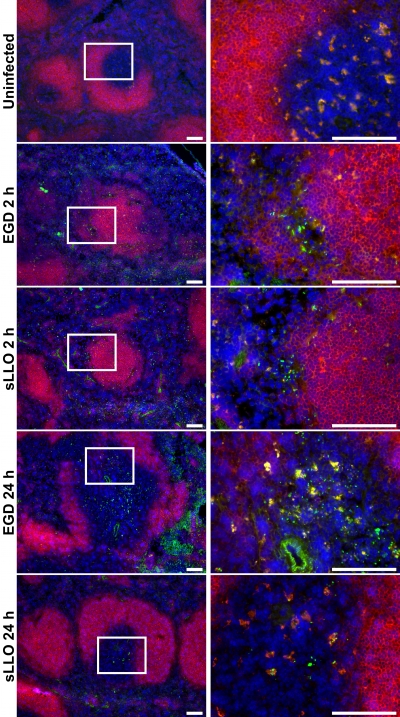
FIG. 4.
Localization of the sLLO and EGD strain cells in infected spleens. Mice were either left uninfected or were infected with the EGD or sLLO strain for the times indicated. For the 2-h infections, mice received 108 CFU of either the EGD or sLLO strain. For the 24-h infections, mice received 106 CFU of the EGD strain or 107 CFU of the sLLO strain. Spleens were isolated from all mice, flash frozen, sectioned, and stained against L. monocytogenes (green), CD19 (red), or DAPI (4′,6-diamidino-2-phenylindole; blue). The white insert box in the left panels indicates the region shown on the corresponding right panels. White bars represent 100 μm. Fields are representative of at least three mice per group.

