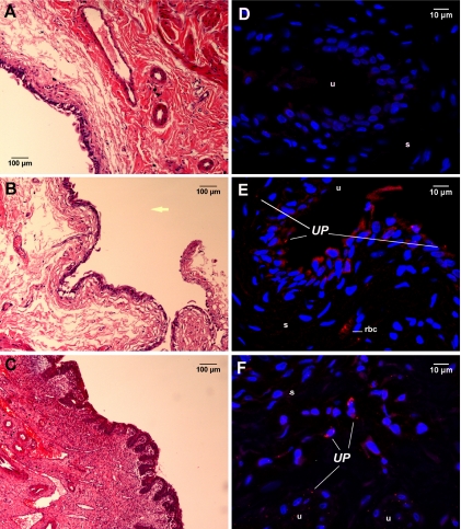
FIG. 2.
Histopathologic profiles and in situ location of U. parvum in the bladder tissues of F344 rats experimentally infected for 2 weeks. (A to C) Images of hematoxylin-eosin-stained sections of bladder tissues from control animals (A) and animals with asymptomatic UTI (B) and complicated UTI (C) were taken at a magnification of ×200. The yellow arrow is placed in the area that would represent the bladder lumen. (D to F) Immunofluorescent images of tissues from uninfected control animals (D) and animals with asymptomatic UTI (E) and complicated UTI (F) were taken at a magnification of ×600; images are 2D projections of image stacks. U. parvum (UP) was labeled with Alexa Fluor-594 (red), and nuclei were stained with DAPI (blue). Panel D shows a U. parvum-positive bladder section stained with isotype antibody, therefore demonstrating the degrees of nonspecific binding and tissue autofluorescence. s, submucosa; u, uroepithelium; rbc, red blood cell.

