FIG. 1.
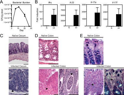
C. rodentium infection in mice. (A) Kinetics of C. rodentium colonization of adult C57BL/6 mice. The C. rodentium burden per gram of stool was measured by plating for nalidixic acid-resistant colonies after infection with C. rodentium containing a nalidixic acid resistance marker. No nalidixic acid-resistant colonies were detected in the absence of C. rodentium infection. (B) Cytokine expression on day 8 as assessed by real-time PCR in intestinal samples from naive (N) and C. rodentium-infected (Inf) mice. The results represent the fold induction over naive WT mice ± the standard error of the mean. (C) Photomicrographs of H&E-stained sections of cecum obtained from naive and C. rodentium-infected mice at day 13. An image of an infected cecum demonstrates a diminution crypt elongation, mononuclear infiltrates (arrow), and an activated epithelium accompanying infection compared to the naive image. (D) Photomicrographs of H&E-stained sections of colon obtained from naive and C. rodentium-infected mice at day 14. A low-magnification image of the infected colon (left) shows focal ulceration of the mucosa (arrow), crypt elongation, and an inflammatory infiltrate composed predominantly of neutrophils. In the high-magnification image of the infected colon (right), crypt abscesses are focally present (arrow) and the epithelium remains reactive. Bar, 50 μm (all images). (E) Periodic acid-Schiff staining shows altered architecture in an infected colon.

