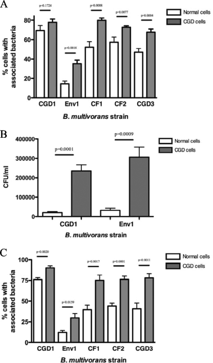
FIG. 3.
Cell association of bacteria with normal and CGD leukocytes. Shown are the associations of five B. multivorans strains with normal and CGD PBMCs (A) and neutrophils (C) and CFU of B. multivorans associated with normal or CGD PBMCs (B). Cell monolayers on coverslips were exposed to bacteria for 2 h (MOI = 4). Coverslips were washed extensively and either stained with Giemsa reagent (A and C) or treated with Triton X-100 and serially diluted with HBSS for microbial cultures (B). Stained coverslips were examined at a ×400 and ×1,000 magnification under light microscopy to determine the percentage of cells with associated bacteria. Quantitative microbial cultures were performed on TSA with 5% sheep blood plates.

