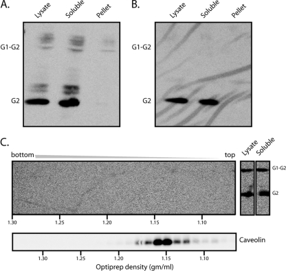
FIG. 2.
GPC segregates with the TX-100-soluble membrane in Candid#1-infected cells and virions. (A) Cells infected with Candid#1 were lysed in cold buffer containing 1% TX-100, and insoluble membranes were separated by high-speed centrifugation. Equal aliquots of the total lysate as well as of the high-speed supernatant (soluble) and the insoluble membrane pellet were incubated with PNGase F and subjected to Western blot analysis using the G2-specific MAb F106G3. Appropriate segregation of detergent-soluble (TfR) and detergent-insoluble (caveolin) cell markers was confirmed in other studies. (B) Candid#1 virions were purified from the cell culture medium by ultracentrifugation through a buffer containing 20% sucrose and were lysed in cold buffer containing 1% TX-100. Equal aliquots of the virion lysate and the detergent-soluble and -insoluble membrane fractions were deglycosylated and analyzed as described for panel A. The G1-G2 precursor and the mature G2 subunit are indicated. Incomplete deglycosylation products from cell lysates are also visible. (C) (Top) The detergent-insoluble fraction from cell lysates prepared as described for panel A was resuspended in TX-100-containing lysis buffer and subjected to OptiPrep density gradient ultracentrifugation in order to float detergent-resistant membranes. Fractions from the resulting gradient (left), as well as aliquots of the total-cell lysate and the high-speed supernatant containing detergent-soluble membranes (right), were analyzed as described for panel A. The positions of G2 and the G1-G2 precursor are indicated. (Bottom) Caveolin-containing lipid rafts were detected in parallel gradients by Western blot analysis. Buoyant densities calculated from the refractive indices of fractions (44) are plotted along the horizontal axes to standardize individual gradients.

