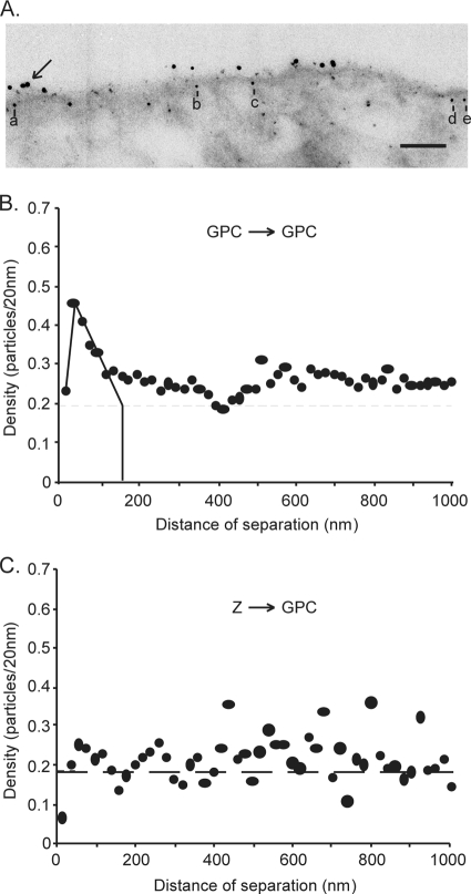
FIG. 4.
Analysis of the organization of GPC in the presence of Z. Vero cells were cotransfected with plasmids encoding GPC and Spep-tagged Z. Cells were fixed, permeabilized, and double-labeled first with the mouse anti-G1 MAb BE08 and biotinylated S protein and then with a secondary antibody conjugated with 12-nm-diameter colloidal gold particles and streptavidin conjugated with 6-nm-diameter colloidal gold particles. (A) Representative electron micrograph of cells double-labeled for GPC and Z. The arrow indicates a cluster of 12-nm-diameter gold particles, and the letters a to e indicate 6-nm-diameter gold particles within 30 nm of the trace of the plasma membrane, which were judged to be associated with the membrane itself. Bar, 100 nm. (B) Pairwise distances between all 12-nm-diameter gold particles (GPC), analyzed as described for Fig. 3. (C) Measurements were collected between each 6-nm-diameter gold particle (Z) within 30 nm of the trace of the plasma membrane and every 12-nm-diameter gold particle (GPC). The density was calculated as the number of 12-nm-diameter gold particles in 20-nm distance increments from each 6-nm-diameter gold particle and was normalized to the number of gold particles analyzed. The normalized density of 12-nm-diameter particles/20 nm was plotted on the y axis against the distance from the 6-nm-diameter particles on the x axis in a histogram. The average density of GPC labeling in the plasma membrane is indicated by the horizontal dashed line.

