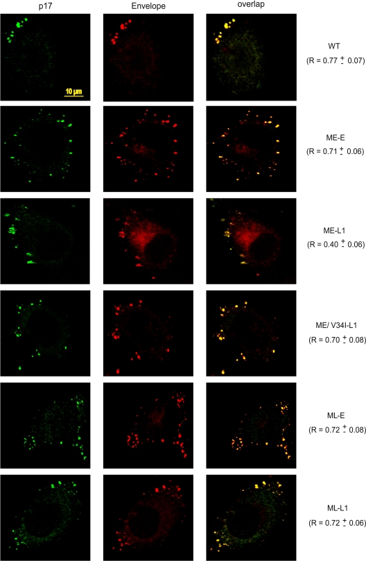
FIG. 6.
Analysis of Gag and Env intracellular distributions by confocal microscopy. HeLa cells transfected with the indicated molecular clones were fixed and processed for immunofluorescence analysis with 2G12 anti-Env MAb and a mouse MAb specific for the matrix p17 protein. Before fixation, all of the cells were treated with 50 μg of cycloheximide/ml for 3 h. Protein distribution was assessed by confocal microscopy and the Pearson channel correlation coefficient (R) (“1” indicating perfect colocalization, “0” indicating no correlation) was calculated for each sample, as described in Materials and Methods. Scale bar, 10 μm.

