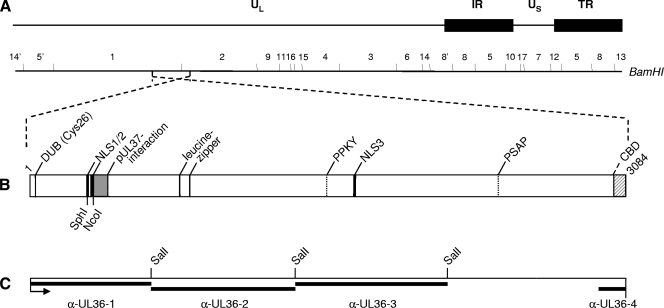
FIG. 1.
Schematic overview of PrV pUL36. (A) Diagram of the PrV genome divided into unique long (UL) and unique short (US) regions by internal repeats (IR) and terminal repeats (TR). The positions of BamHI restriction sites are indicated. (B) Confirmed and putative functional domains in PrV pUL36 are indicated, such as DUB (Cys26), the active-site cysteine of the deubiquitinating activity (28); the pUL37 interacting domain (15, 31); the NLS; the leucine zipper; putative late domain motifs (PPKY and PSAP) (4); and CBD, the capsid binding domain (8). SphI and NcoI sites used for deletion of the two NLS motifs are also indicated. (C) Regions of pUL36 contained in GST-expression proteins used for immunization are marked by black bars above the designations of the resulting antisera. α-, anti-. The SalI restriction sites used for cloning of pGEX-UL36-2 and pGEX-UL36-3 are indicated.

