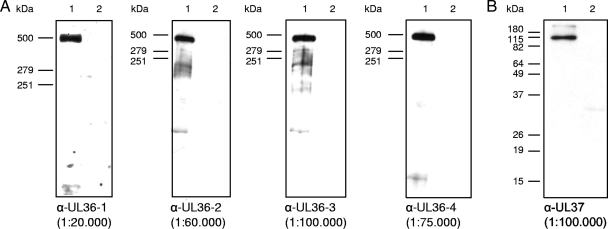
FIG. 2.
Western blot analyses of purified PrV virions. Purified virions of PrV-Ka (lanes 1) and lysates of noninfected RK13 cells (lanes 2) were separated on 6% (A) or 10% (B) polyacrylamide gels. α-, anti-. After electrotransfer onto nitrocellulose membranes, parallel blots were incubated with the indicated dilutions of the four antisera directed against PrV pUL36 (A). For a control, an antiserum against pUL37 was included (B). Locations of molecular marker proteins (high molecular mass marker; Invitrogen) are indicated to the left of each panel.

