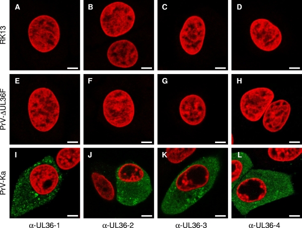
FIG. 3.
Indirect immunofluorescence of infected cells. Indirect immunofluorescence of RK13 cells, either infected with PrV-Ka or PrV-ΔUL36F for 24 h or mock infected, with each of the four anti-pUL36 antisera is shown in green. Chromatin was counterstained with propidium iodide or ToPro-3 (shown in red). α-, anti-. Scale bars represent 5 μm.

