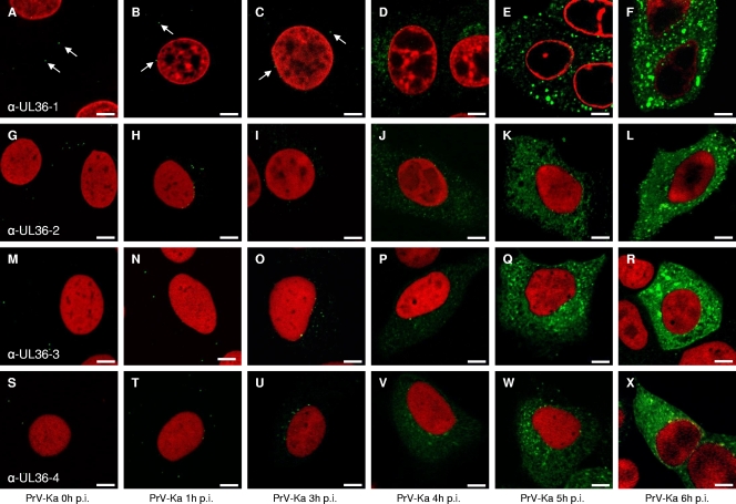
FIG. 4.
Intracellular localization of pUL36 during PrV infection. RK13 cells were infected synchronously with PrV-Ka at an MOI of 5 and fixed 0, 1, 3, 4, 5, and 6 h postinfection (p.i.). Immunofluorescence was performed by confocal laser-scanning microscopy using anti-UL36-1 (top row), anti-UL36-2 (second row), anti-UL36-3 (third row), and anti-UL36-4 (bottom row) antisera. Chromatin was counterstained with propidium iodide or ToPro-3 (shown in red). Arrows point to fluorescent dots most likely representing virus particles. α-, anti-. Scale bars represent 5 μm.

