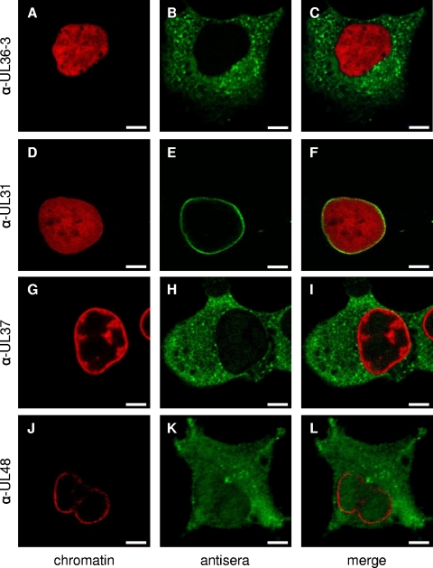
FIG. 5.
Indirect immunofluorescence of PrV-infected cells with antisera directed against pUL36, pUL31, pUL37, and pUL48. Cells were infected synchronously with PrV-Ka and fixed after 5 h. Immunofluorescence analysis was performed by confocal laser-scanning microscopy using anti-UL36-3 (top row), anti-UL31 (second row), anti-UL37 (third row), and anti-UL48 (bottom row) antisera and Alexa 488-conjugated secondary antibodies (green). Chromatin was counterstained with ToPro-3 or propidium iodide (shown in red). α-, anti-. Scale bars represent 5 μm.

