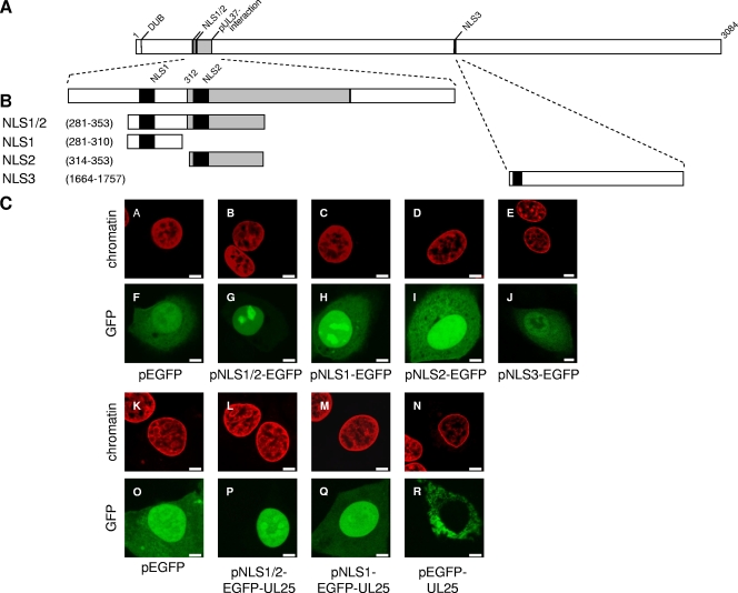
FIG. 6.
Nuclear targeting properties of predicted NLS motifs. (A) Schematic overview of PrV pUL36. Panel B shows enlarged views of the N-terminal region containing the putative NLS motifs NLS1 (aa 288 to 296) and NLS2 (aa 315 to 321) and the C-terminal segment containing NLS3 (aa 1679 to 1682). NLS are shown as black bars, and the pUL37-interacting domain is shaded in gray. Fragments of PrV pUL36 containing putative NLS motifs were fused in frame to the 5′ end of the EGFP- or EGFP-UL25 genes (not shown). Numbers indicate amino acid positions in PrV pUL36. DUB denotes the active-site cysteine of the deubiquitinating activity (28). (C) Intracellular localization of NLS-EGFP or NLS-EGFP-UL25 fusion proteins in RK13 cells. GFP autofluorescence (green) and ToPro-3-stained chromatin (red) were recorded in a confocal laser-scanning microscope. Scale bars represent 5 μm.

