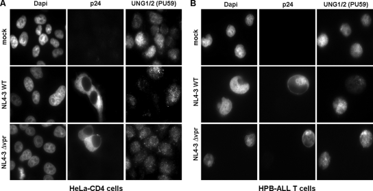
FIG. 2.
Immunofluorescence analysis of UNG2 downregulation in HIV-1-infected cells. HeLa-CD4 (A) or HPB-ALL T (B) cells were infected with wild type (middle panels) or Δvpr (lower panels) HIV-1NL43 and were then analyzed 48 h after infection by immunofluorescence. Cells were fixed, permeabilized, and subsequently stained with anti-UNG (PU59), anti-p24 antibodies and DAPI (4′,6′-diamidino-2-phenylindole). Cells were analyzed by epifluorescence microscopy, and images were acquired by using a charge-coupled device camera. WT, wild type.

