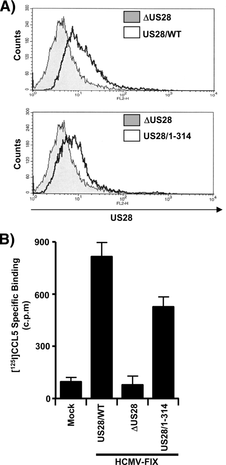
FIG. 3.
US28/WT and US28/1-314 proteins exhibit similar cell surface expression and ligand-binding activity in infected cells. (A) HFFs were infected with HCMV ΔUS28 or HCMV FLAG-US28/WT (top panel) and with HCMVΔUS28 or HCMV FLAG-US28/1-314 (bottom panel) viruses for 48 h. Surface expression of FLAG-US28 on infected cells was detected by staining with FLAG-specific M2-biotin, followed by streptavidin-PE and analyzed by FACS. The histograms shown are representative of six independent experiments performed in duplicate. (B) Infected HFFs were incubated with 28 pM [125I]CCL5 in the absence or presence of 14 nM unlabeled RANTES to discriminate between specific and nonspecific binding. The data shown represent specific binding of [125I]CCL5, as assessed by liquid scintillation chromatography, and are derived from six independent experiments performed in duplicate and represent the means ± the standard errors of the mean (SEM).

